Advanced 3D Printing for Surgical Planning
Case-Specific 3D Anatomical Models
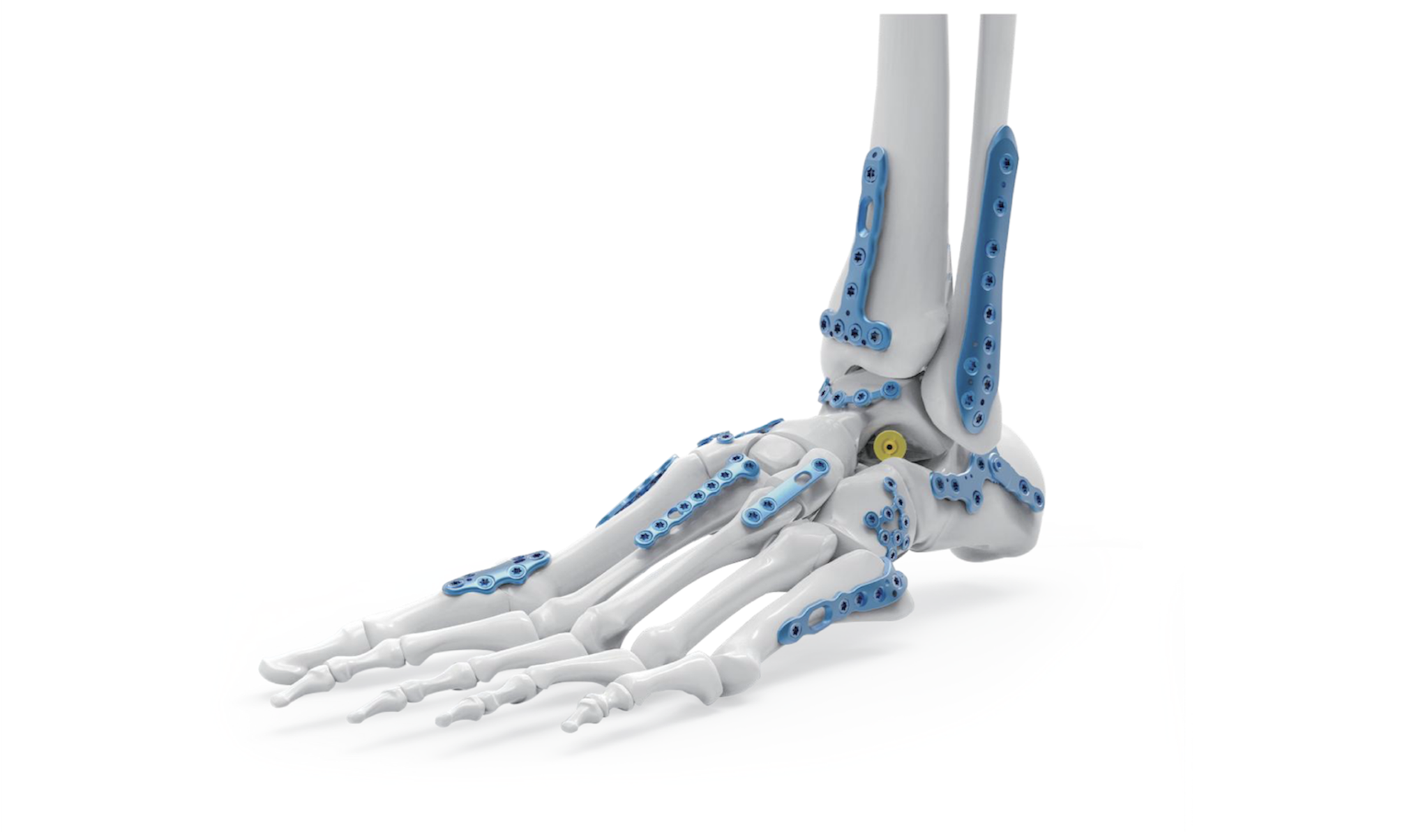
Alongside the distribution of orthopaedic implant systems, we support preoperative planning and implant selection with 3D-printed patient-specific anatomical models.
Based on CT DICOM data provided for the case, we can segment the relevant anatomical structures and convert them into accurate digital 3D models. These models help visualise complex anatomy and provide a clear physical reference during preoperative preparation.
DICOM Data
The workflow begins with medical imaging data, typically acquired through a high-resolution CT scan and provided in DICOM format. The DICOM image series contains the detailed anatomical information required for the creation of the patient-specific anatomical models.
The imaging dataset is reviewed to assess image quality, resolution and the visibility of the anatomical structures relevant to the individual case. Depending on the clinical objective, specific regions of interest can then be selected for further processing, such as bones, joints, fracture fragments or other anatomical structures.
The prepared DICOM dataset provides the foundation for the subsequent segmentation and 3D reconstruction process, allowing the relevant patient anatomy to be converted from two-dimensional medical images into an accurate digital 3D representation.
Segmentation
During the segmentation process, the anatomical structures relevant to the individual case are identified and separated from the surrounding tissues within the CT dataset. This step converts the medical imaging data into a basis of 3D printing for surgical planning, creating clearly defined anatomical regions that can subsequently be reconstructed as three-dimensional digital models.
Depending on the purpose of the case, individual bones, joints, fracture fragments or other structures can be segmented separately. In complex trauma or anatomical deformities, separate segmentation can provide a clearer understanding of the spatial relationship between individual anatomical components.
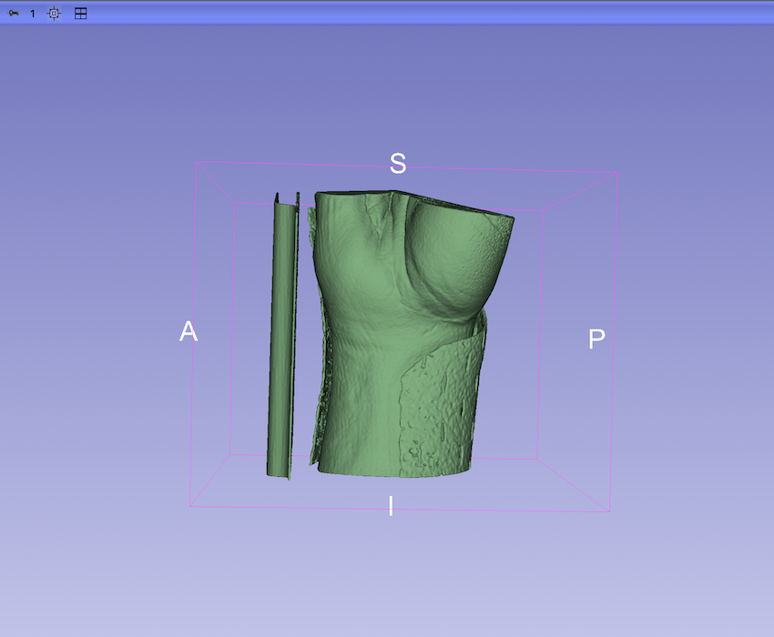
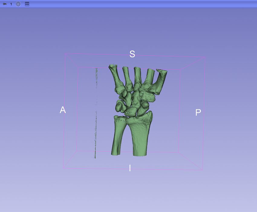
The segmented anatomy is carefully reviewed and refined to reduce imaging artefacts and preserve clinically relevant anatomical details. Once the segmentation is completed, the resulting data provides the basis for the creation of an accurate patient-specific digital 3D model suitable for further anatomical assessment, surgical planning and physical model production.
3D Modelling
Following segmentation, the selected anatomical structures are converted into a patient-specific digital 3D model. The reconstructed geometry represents the relevant anatomy in three dimensions, allowing complex anatomical relationships to be examined from different perspectives beyond conventional two-dimensional CT images.
The digital model is carefully reviewed and refined where necessary. Minor artefacts originating from the imaging or segmentation process can be corrected, while clinically relevant anatomical features and spatial relationships are preserved.
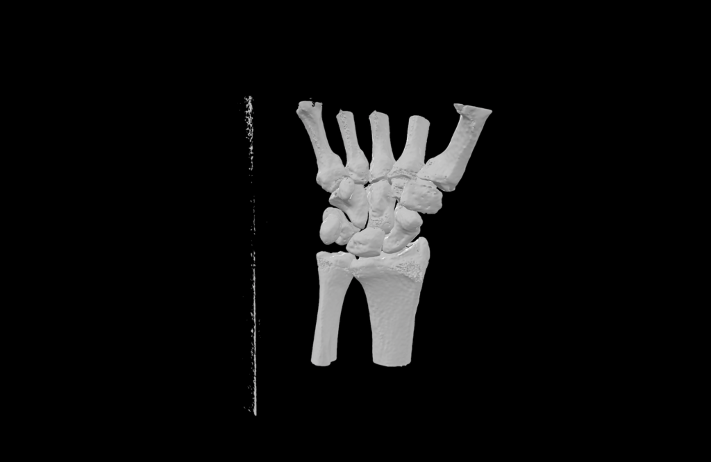
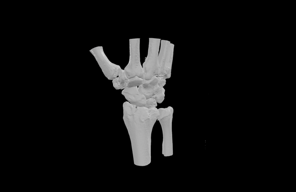
Depending on the requirements of the individual case, anatomical structures or fracture fragments can be prepared as separate components. The completed digital model can then be used for anatomical assessment, preoperative planning, implant positioning or sizing evaluation, and as the basis for manufacturing an accurate physical 3D anatomical model.
Additive Manufacturing
Once the digital 3D model has been prepared and verified, it can be converted into a physical patient-specific anatomical model using additive manufacturing technologies. The manufacturing process is selected according to the anatomical complexity, required level of detail and intended application of the model.
3D printing for surgical planning can result perfect physical model reproduction to the relevant patient anatomy and can be manufactured at true anatomical scale or, when required, enlarged to allow small or complex structures to be examined in greater detail.
Depending on the application, different materials and manufacturing parameters can be selected to achieve the required dimensional accuracy, surface quality and mechanical characteristics. Individual anatomical structures or fracture fragments may also be produced as separate components where this provides additional value during assessment or planning.
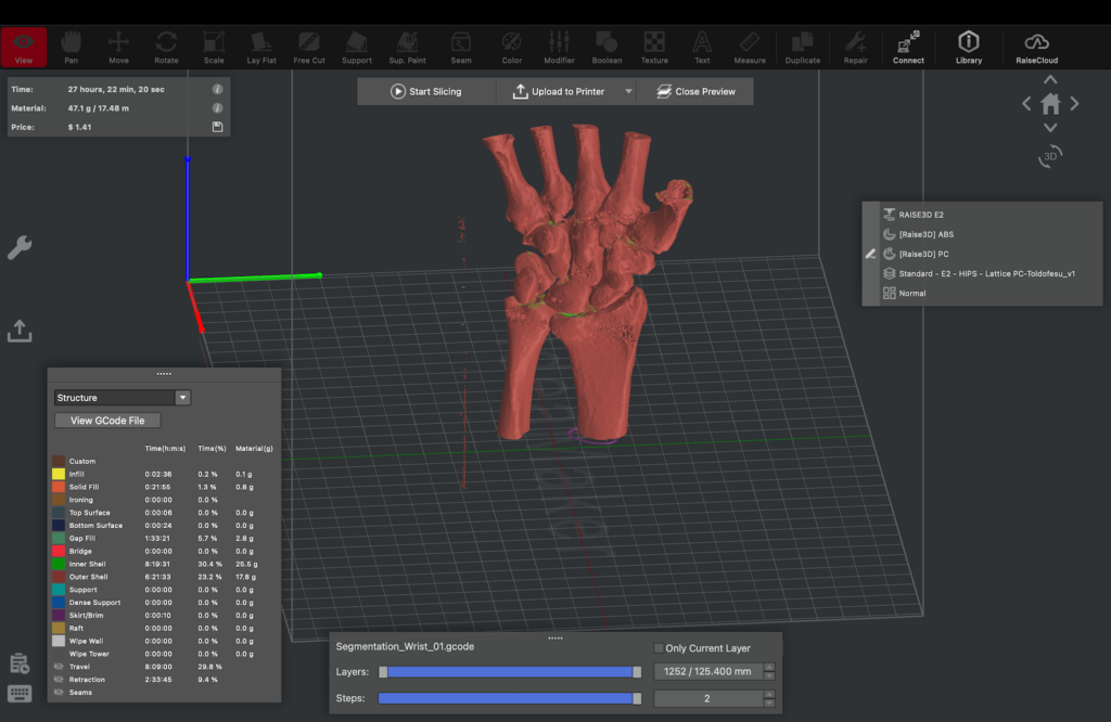
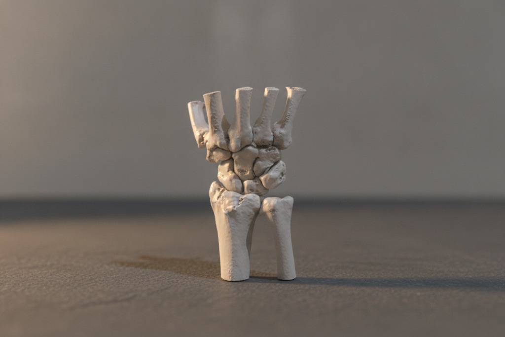
The completed patient-specific anatomical model provides a tangible representation and can support anatomical assessment, preoperative preparation, surgical planning, implant selection and professional communication. 3D printing for surgical planning can support surgeons on new ways, and can help to achieve higher precision and save time.
Surgical Planning
Patient-specific anatomical models, printed in 3D can provide valuable support during preoperative surgical planning by allowing complex anatomy to be examined and evaluated before the procedure.
Unlike conventional two-dimensional CT images, a three-dimensional model provides a direct representation of anatomical relationships, deformities and fracture configurations. Physical models can also be handled and examined from different angles, providing an additional perspective when evaluating complex cases.
3D printing for surgical planning can create the models may support the assessment of surgical approaches, fracture reduction strategies and the spatial relationship between relevant anatomical structures. Where appropriate, different implant sizes, positions or configurations can also be evaluated on the patient-specific anatomy before surgery.
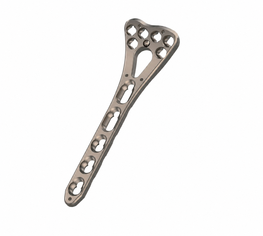
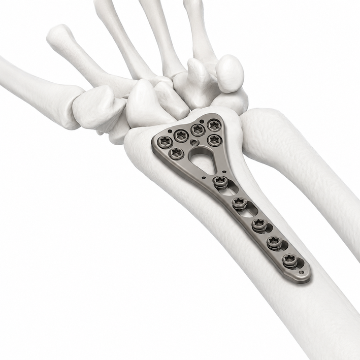
This approach can provide surgeons with additional information during case preparation and may facilitate multidisciplinary discussion, communication and the development of an appropriate surgical strategy for the individual patient.
Medical 3D Print Materials
ABS Medical
ABS Medical is a specialized 3D printing filament made from ABS resin that meets biocompatibility requirements (USP Class VI and ISO 10993), designed for advanced medical and technical applications and for skin contact for up to 30 days; the material is approved for food contact (EU 10/2011, US FDA 21 CFR 181.32) and offers high mechanical and thermal resistance, dimensional stability, and very strong interlayer adhesion, making it suitable for, among others, parts for medical devices, components for pharmaceutical production lines, and rehabilitation elements.
PLA / Medical PLA
PLA Medical is a bio-based, medical-grade polymer composed of high-quality Polylactic Acid [PLA]. This bone white filament can be used to manufacture 3D parts such as patient-specific anatomical models, surgical planning models, prototypes, and other applications that come into contact with the skin. PLA is the base material of 3D printing for surgical planning activities since it represent bone characteristics so precise.
Polylactic acid [PLA] is a semi-crystalline thermoplastic that is bone white in color, rigid, and dimensionally stable. PLA is derived from bio-based granules that comply with ISO 10993-5, enabling the production of 3D-printed parts that come into contact with the skin, such as surgical planning models, medical device prototypes, interfaces, and tools for functional prototyping. It is prefect material for patient-specific anatomical models and 3D printing for surgical planning.
PLA (non Medical version) is having the same mechanical properties as medical grade owns but cannot be contacted with skin for as long time as medical could be. It is perfect for surgical modelling, prototyping and for demonstrational applications like patient-specific anatomical models.
Medical Polycarbonate (PC)
OSSFILA® Medical Polycarbonate Filament is a biocompatible, non-toxic and chemical resistant 3D printing material perfect for printing surgical guides, medical devices and medical equipment. It is an alternative to PMMA with better impact resistance, rigidity and drillability. PC is the perfect material if autoclavable properties are needed.
Features
- High optical transparency
- Outstanding impact resistance polishability & UV tolerance
- Resistant to most chemicals especially acids
- Biocompatible and non-toxic (USP 88 Class VI)
- Sterilizable by gamma radiation, e-beam, autoclave and ethylene oxide
Suggested Applications
Surgical instrument and training models including body fluid container, microfluidic device, pump impeller, stirrers, surgical trays and many more.