3D Printing for Veterinary Applications
Implant Systems, 3D Technology and Veterinary Surgical Planning
We provide veterinary orthopaedic solutions combining high-quality implant systems with advanced 3D printing in veterinary applications and case-specific anatomical modelling. Our goal is to support veterinary surgeons throughout the surgical planning process — from implant selection and preoperative preparation to training and demonstration.
Case-Specific 3D Anatomical Models and Implant Selection Support
Submission of DICOM Data
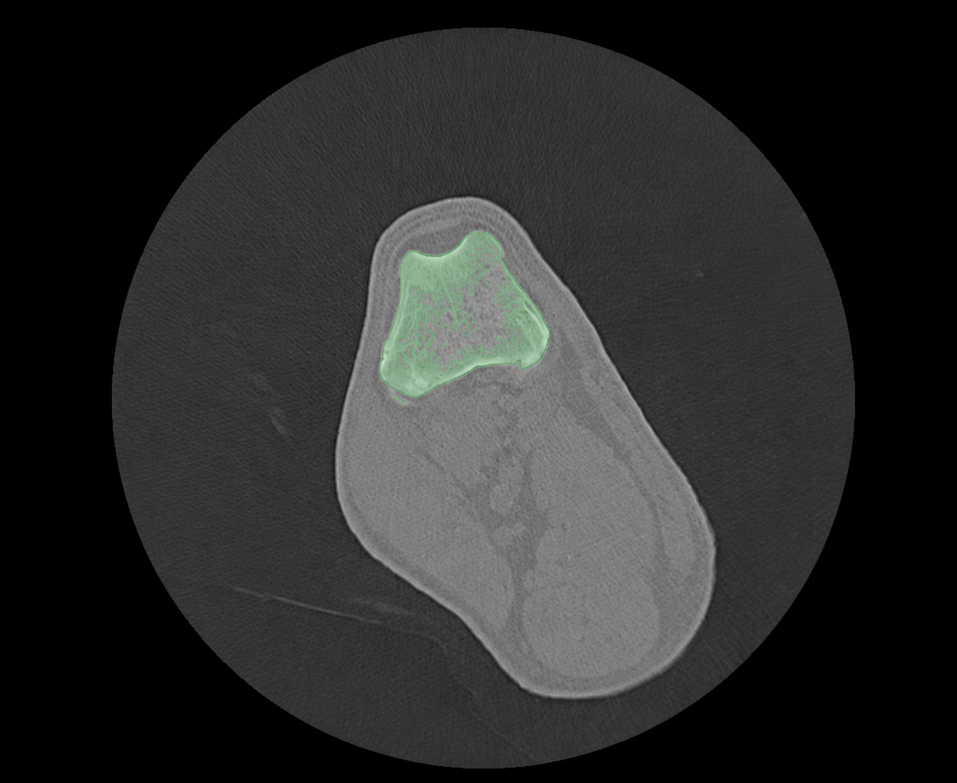
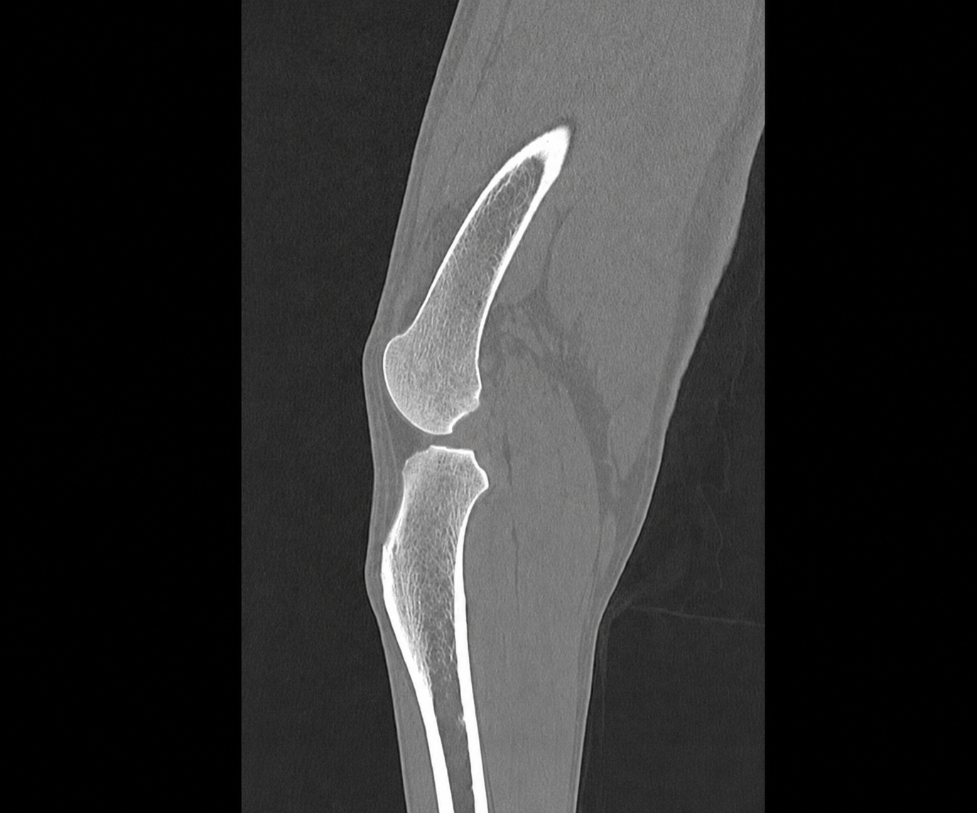
The process begins with the secure transfer of the DICOM data generated during the CT examination. These files contain the detailed radiological information required to reconstruct the relevant anatomical region in three dimensions.
Processing can be performed for a wide range of anatomical regions, including:
- limbs and joints
- pelvis
- spine
- skull and jaw
- radius, ulna, and humerus
- femur and tibia
- as well as other complex bone anatomical structures
For veterinary applications, the same workflow can also be used to support the preparation of TPLO, TTA, ALPS, trauma, orthopaedic, or reconstructive cases. 3D printing for veterinary applications can support this from all way and can achieve precise results in veterinary surgical planning.
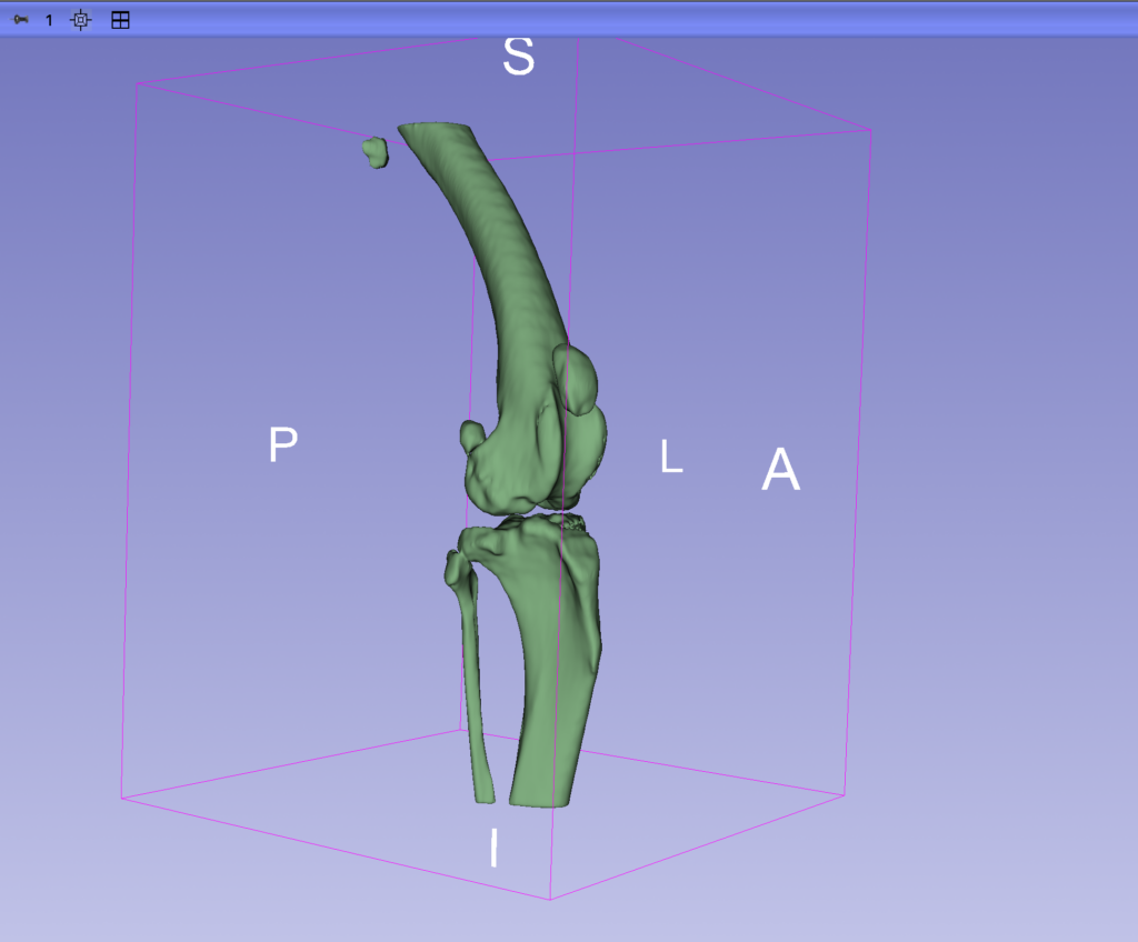
Segmentation and Digital 3D Model Creation
A high-resolution digital 3D model is created from the segmented anatomical data. The data would be the source of the 3D printing of many kinds of veterinary surgical planning applications.
The model can be further optimised for physical manufacturing where required. This may include correcting minor artefacts originating from the imaging process, removing unnecessary structures, or separating the model into individual components.
Physical 3D Anatomical Model
3D printing for veterinary applications results a physical model that can provide valuable support during preoperative preparation because, unlike CT images or digital 3D visualisations displayed on a screen, the anatomy can be examined directly and physically.
The model may be used to assess in advance:
- the expected implant position,
- appropriate plate length,
- possible screw trajectories,
- surgical access,
- the extent of an anatomical deformity,
- and the three-dimensional configuration of a complex fracture.
The digital model can also be used to compare different implant positions or plate sizes before surgery. If required, the anatomical model can be enlarged, typically to 100-400% of its original size, allowing small or complex structures to be examined greater detail during surgical planning.
The models may also be used for educational, demonstration, and communication purposes, for example during multidisciplinary surgical planning or when presenting a complex case.
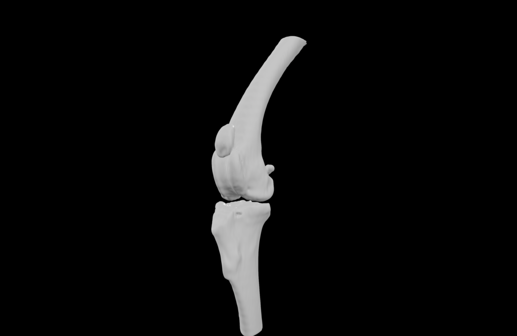
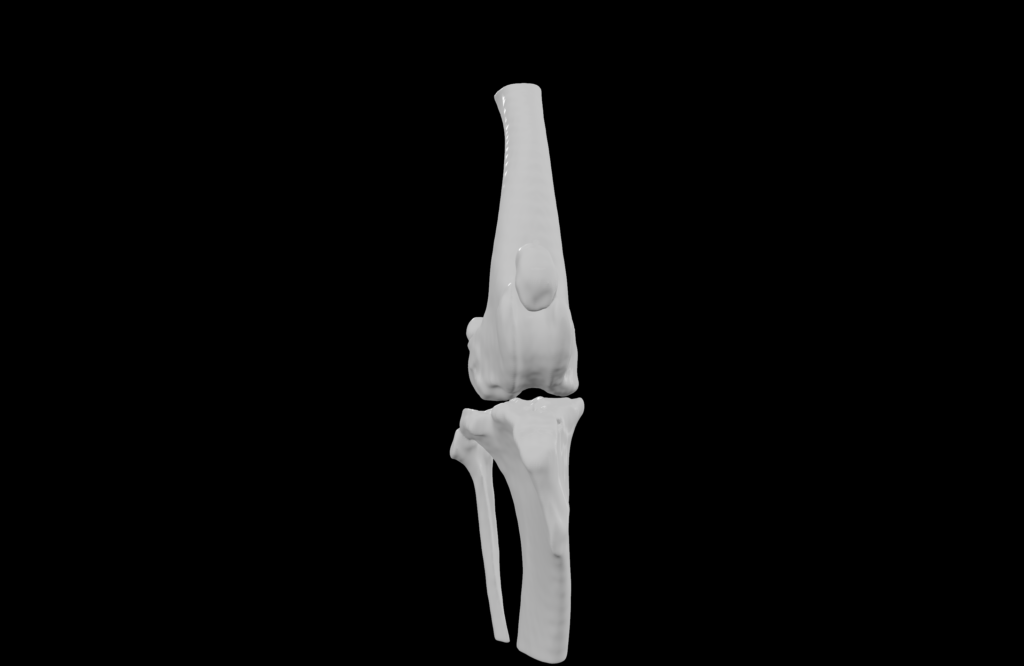
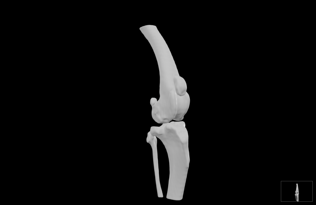
Veterinary Surgical Planning
Patient-specific digital and physical 3D anatomical models and 3D printing for veterinary applications can provide valuable support during preoperative planning in veterinary surgery, particularly 3D printing for veterinary applications can create higher precision and efficiency during the complex orthopaedic, trauma, and reconstructive cases.
Three-dimensional models created from CT data allow detailed assessment of anatomical relationships, deformities, fracture configurations, and the affected bone structures. Compared with conventional two-dimensional imaging, a 3D model provides a more direct understanding of spatial relationships, while a physical model can also be handled and examined from multiple angles.
During surgical planning, the models may support:
- evaluation of the planned surgical approach,
- assessment of fracture fragments and deformities in three dimensions,
- selection and positioning of implants,
- preoperative fitting of plates and other implants,
- planning of osteotomies and corrective procedures,
- preparation for complex TPLO, TTA, trauma, and reconstructive cases.
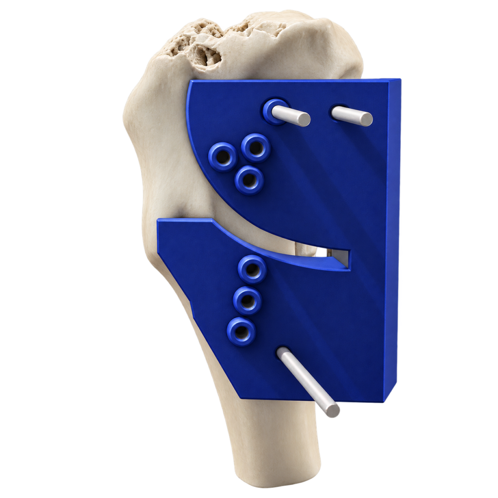
Where required, the digital anatomical model can also serve as the basis for the design of patient-specific surgical aids, such as drill guides or cutting guides.
The aim is to provide the surgeon with a clearer understanding of the individual anatomy before the procedure and to support the evaluation of different options when developing the most appropriate surgical strategy for the specific case.
Fracture Reduction Guides
Patient-specific fracture reduction guides can support the accurate repositioning and alignment of bone fragments during the surgical treatment of complex fractures.
Based on CT imaging and the segmented 3D anatomy, the fracture can be virtually reconstructed and the desired anatomical alignment planned before surgery. A patient-specific guide can then be designed to correspond to the individual bone geometry and the planned reduction.
Fracture reduction guides may support:
- repositioning of displaced bone fragments,
- restoration of anatomical alignment,
- control of rotational and angular correction,
- maintenance of the planned reduction during fixation,
- positioning of plates, screws, or other fixation devices,
- preparation for complex trauma and reconstructive procedures.
Where appropriate, reduction guides may also be combined with patient-specific drill or cutting guides as part of a complete surgical planning workflow.
The objective is to transfer the preoperative digital plan into the surgical procedure in a controlled and reproducible way, providing additional support in cases where fracture geometry or anatomical deformity makes conventional reduction more challenging.
Tibial Plateau Leveling Ostheotomy Guides
Patient-specific 3D planning for veterinary applications can provide additional support during Tibial Plateau Leveling Osteotomy (TPLO), particularly in cases involving complex anatomy, deformity, previous surgery, or unusual tibial geometry.
Using CT imaging and a segmented 3D model of the tibia, the procedure can be planned virtually before surgery. The digital model allows assessment of the proximal tibial anatomy, tibial plateau orientation, osteotomy position, and the spatial relationship of the relevant anatomical structures.
Patient-specific TPLO planning may support:
- evaluation of tibial anatomy and deformity,
- planning of the osteotomy position and orientation,
- assessment of the intended tibial plateau rotation,
- preoperative evaluation of plate size and positioning,
- planning of screw trajectories,
- improved transfer of the digital surgical plan to the operating room.
Where required, a patient-specific cutting and/or drill guide can be designed to fit the individual tibial anatomy and reproduce the planned osteotomy and fixation positions during surgery.
The aim is to provide the surgeon with a case-specific planning tool that supports accurate preparation and more consistent execution of the planned TPLO procedure. 3D printing for veterinary applications can create high value and improvement in many areas of the field.